
Imaging & AI
Before AI Can Read the Plaque, Someone Has to Fix the Image
Motion correction can improve coronary CT interpretation. Proving that it also improves automated plaque measurements requires a different kind of validation.
Significance 4/10: Motion correction improves coronary CT images, but its effect on plaque measurement is untested.
Coverage we found: no other coverage since the Morikawa study was published in 2026 (searched October 13, 2026).
How we scored this
- Strength of evidenceTwo retrospective single-workflow studies, 279 and 151 patients.
- Effect on practice or understandingInforms how teams should validate plaque software on motion-corrected images.
- ReachRelevant to coronary CT labs using this reconstruction and plaque analysis.
- StakesIndirect: scans evaluable without beta-blockers, and measurement reliability.
- SurpriseThat cleaner images help is expected; the article's point is what has not been tested.
Coverage found
We found no other coverage in our searches.
Only news, trade and journal coverage is counted. Press releases and other sources, such as reference sites, blogs, social posts and stock-news reprints, are listed but not counted. Searches miss things: tell us about coverage we missed. How the Unread Index works.
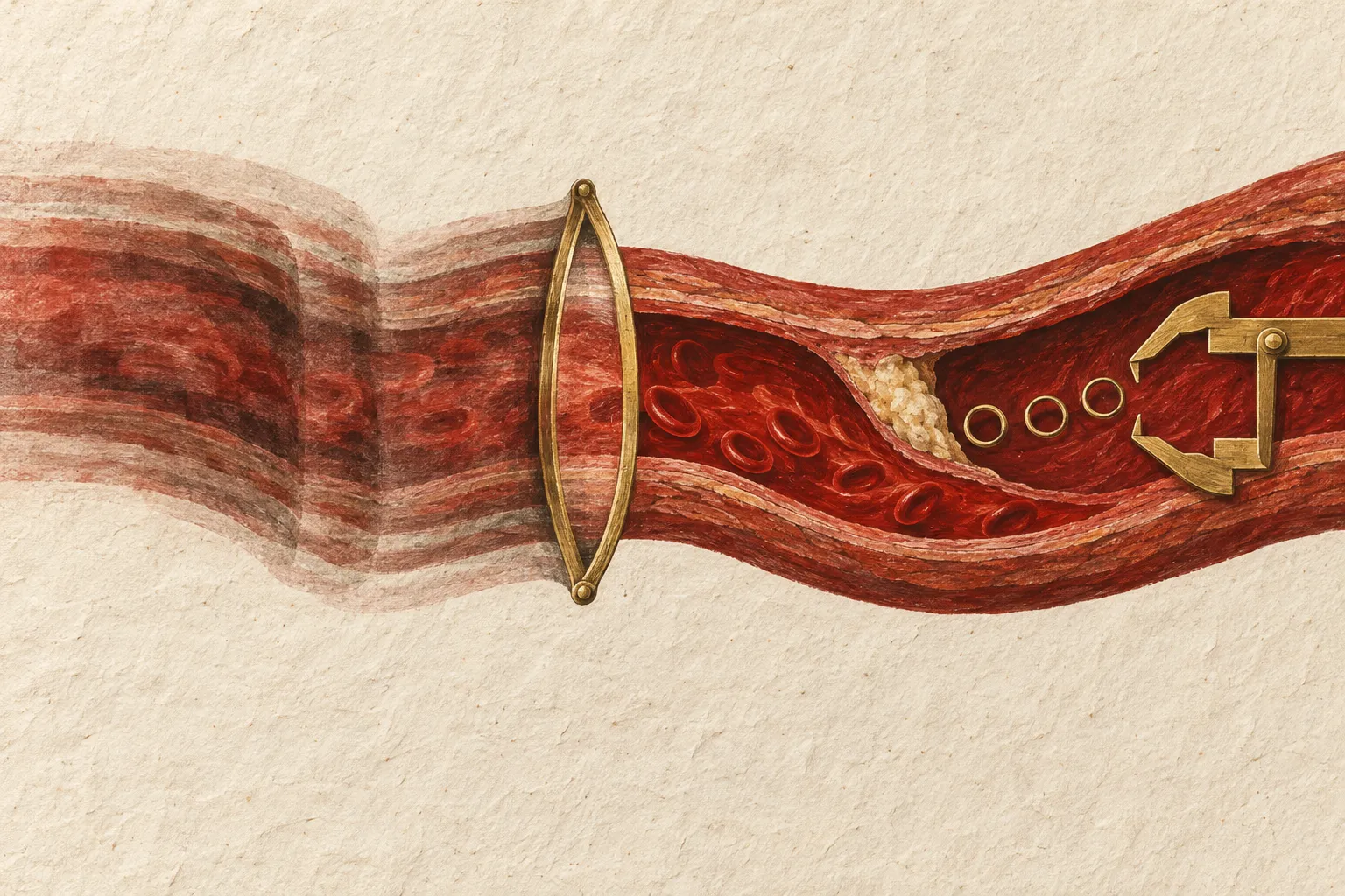
A cleaner coronary CT image looks like an obvious head start for automated plaque analysis. It’s also easy to treat that head start as the finish line. Showing that coronary CT motion correction reduces artifacts doesn’t show that a separate program measures plaque more accurately from the corrected images.
Two studies of second-generation SnapShot Freeze motion correction (SSF2) mark where the evidence currently stops. One looks at image quality and phase selection. The other tests a downstream physiology calculation. Neither directly validates automated measurement of plaque composition or volume.
Coronary CT motion correction and phase selection
In a 2026 retrospective study, Tomoro Morikawa and colleagues looked at coronary CT angiography done without beta-blockers on a 256-row scanner, comparing systolic and diastolic reconstructions with and without SSF2.
The abstract lists 368 patients in the methods but reports results for 279, without explaining the difference. Among those reported, heart-rate-guided phase selection with a cutoff of 67 beats per minute made 91.8% of scans evaluable. In the 50 patients who also had invasive coronary angiography, accuracy for detecting stenosis of 50% or more was 89.0%.
Those numbers describe one acquisition and reconstruction workflow. They don’t tell every scanner, patient population or analysis package to use the same cutoff, and they don’t make the case for dropping beta-blockers across coronary CT practice.
A downstream result, with a different endpoint
A 2024 study by Wenli Yang and colleagues gets closer to what happens after reconstruction. It retrospectively compared standard images, first-generation motion correction and SSF2 in 151 patients who had coronary CT plus invasive coronary angiography or fractional flow reserve assessment within three months.
SSF2 scored better on image quality. At the lesion level, CT-derived fractional flow reserve from SSF2 images reached an area under the curve of 0.887 for identifying ischemic lesions under the study’s definition, against 0.795 with first-generation correction.
So reconstruction can move a downstream analytic result. But the endpoint here was CT-derived fractional flow reserve and stenosis. Plaque quantification asks something else: how much material sits in the vessel wall, and what kind.
The missing plaque experiment
What these studies don’t show is whether coronary CT motion correction improves a specific plaque-analysis system’s agreement with a suitable reference standard, or makes repeat measurements more consistent.
A convincing test would name the scanner, reconstruction settings and exact software version. It would compare corrected and uncorrected images from the same exams, report which scans couldn’t be analyzed, and check whether any gain holds up across heart rates and levels of image quality. Independent validation would count for more than a good-looking example image.
It would also keep measurement agreement separate from clinical usefulness. A more reproducible plaque number may be technically valuable. Whether it changes management or predicts outcomes better needs its own evidence.
What to ask before adopting the workflow
For imaging teams weighing coronary CT motion correction ahead of plaque analysis, the vendor conversation should be specific. Which input series is the plaque software validated to accept? Was motion-corrected reconstruction part of that validation? Can the lab tell when correction helped interpretation and when a study is still unfit for quantitative analysis?
Neither study ties SSF2 to a particular commercial plaque platform or shows better plaque measurements from that pairing.
The research makes a fair case for taking reconstruction seriously. The next step is testing the whole measurement chain. A cleaner-looking artery is a good input. The plaque numbers still have to earn their credibility.
Edited by a healthcare imaging professional with more than 25 years of experience in the field. How we report and edit.
Read next